HOW IT WORKS
X-ray radiography passes ionizing radiation through a part onto a detector or film on the opposite side. Denser material absorbs more radiation, so a void, inclusion, or wall-thickness variation shows up as a contrast difference in the resulting image — a visual map of internal density rather than a point-by-point echo reading.
Where ultrasonic testing excels at precise depth location, X-ray excels at visualizing complex internal geometry and porosity distribution across a wider area in a single image — which is why the two technologies are often specified together on critical components.
WHY X-RAY
When density, not depth, is the question
Porosity Mapping
Visualizes the distribution and density of trapped gas across castings and welds.
Complex Internal Geometry
Reveals internal structure in parts too geometrically complex for a straightforward UT scan path.
Weld Inspection
A standard method for verifying weld integrity in pressure-critical assemblies.
COMMON QUESTIONS
Should I choose X-ray or ultrasonic testing?
It depends on the flaw type and part geometry. Many critical components use both methods together for complementary coverage — talk to SDI about your specific inspection requirements.
What safety measures does X-ray inspection require?
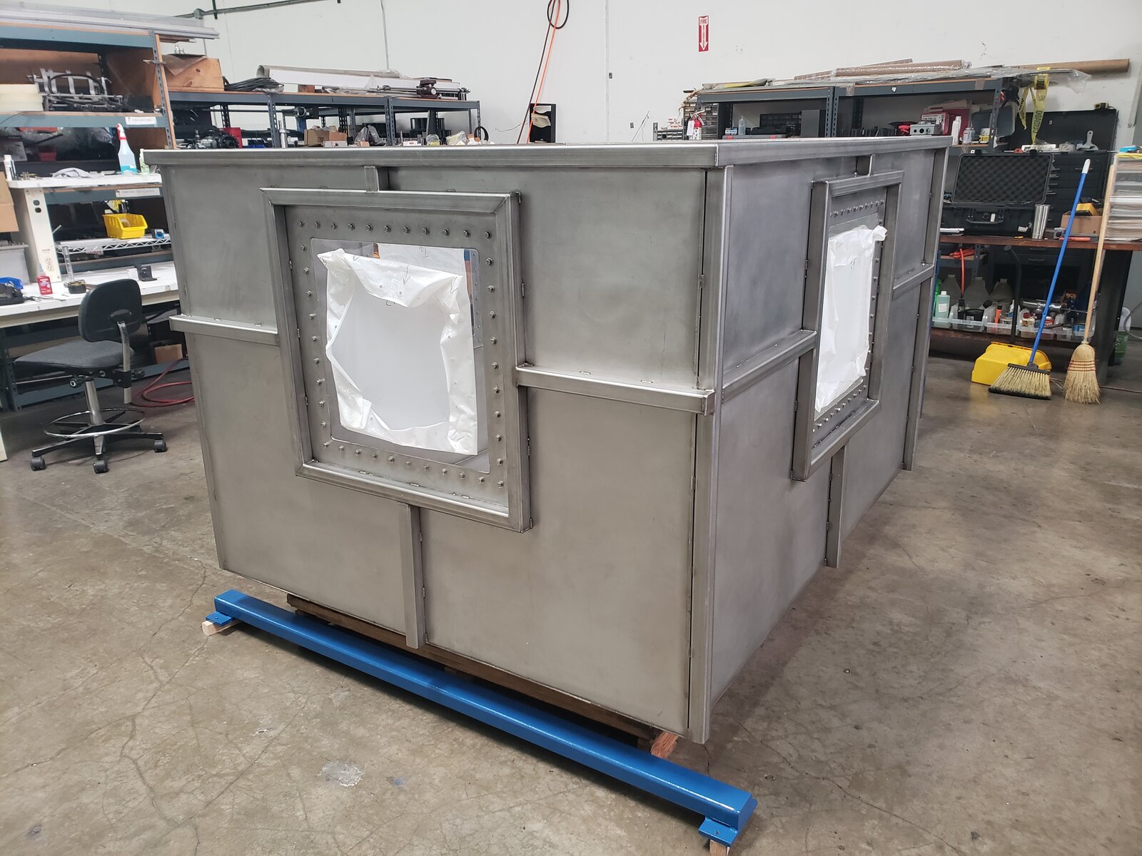
Radiographic inspection requires shielded environments and certified operators, consistent with applicable radiation safety regulations.
Can X-ray inspection be automated like SDI's UT systems?
Automated radiographic inspection is available for production environments — contact SDI to discuss your throughput and part requirements.
Not sure which inspection method fits your part?
SDI's engineers can help you compare technologies.